You found a bump on your horse and opened Google Images — now you’re more confused than before. That’s because equine sarcoid pictures online often lack context, showing advanced cases without clear labels or stages. As a horse owner who’s dealt with these myself, I know the frustration. Sarcoids are the most common skin tumor in horses, affecting up to 12% of the population in some areas. They aren’t cancerous in the spreading sense, but they can grow aggressively and recur if ignored. This visual identification guide cuts through the noise. Below you’ll find clear equine sarcoid images and descriptions of every sarcoid type, organized by appearance and location. We’ll cover what each looks like, where they pop up, and what they get mistaken for. Plus, because early detection matters, we’ll discuss when to call your vet. Remember, these growths stem from bovine papillomavirus, often triggered by minor skin trauma. Use this as your go-to reference to spot issues fast and keep your horse comfortable.
How to Use This Identification Guide
Compare your horse’s growth to the equine sarcoid pictures below. Look closely at size, texture, color, and any ulceration. Note the location — sarcoids favor limbs, head, and underbelly. However, sarcoids can vary by stage, so a small bump today might evolve tomorrow. Always confirm with a vet; visual ID alone isn’t a diagnosis. A biopsy often verifies it’s a sarcoid, ruling out look-alikes like warts or melanoma. As a result, you’ll avoid unnecessary worry or delayed treatment. Meanwhile, document changes with photos over time. This guide empowers you to act early, but professional advice ensures accuracy.
The 6 Types With Pictures
Sarcoids come in six main types, each with distinct looks and behaviors. They all share viral roots but differ in aggression and response to treatment. Here’s a breakdown, complete with equine sarcoid images for easy comparison.
Occult Sarcoid — Pictures & Identification
Occult sarcoids are the sneakiest type, often mistaken for harmless bald patches. They appear as flat, circular areas of hair loss with gray, thickened skin and mild scaling. No raised edges or warts yet — just subtle changes in hair texture or color. These start small, around 1-2 inches, and can stay dormant for years.
Sarcoids | School of Veterinary Medicine
Occult sarcoid on horse neck — equine sarcoid pictures identification.
They typically show up on the face, neck, or inner thighs, where flies or tack rub. Horse owners confuse them with ringworm or rub marks from gear. However, unlike ringworm, they don’t itch or spread quickly. If traumatized, they can morph into more aggressive types like verrucous or nodular. Early spotting prevents that shift.
Sarcoids | School of Veterinary Medicine
Occult sarcoid on horse face — equine sarcoid pictures.
Because they’re mild, many go unnoticed until they change. Check thin-skinned areas regularly.
Verrucous Sarcoid — Pictures & Identification
Verrucous sarcoids resemble warts or scabs, earning their “warty” name. They feature gray, rough, scaly surfaces that may coalesce into larger patches. Small nodules hide beneath, giving a lichen-like texture. These grow slowly and stay flat, rarely ulcerating unless irritated.
Common spots include the body, groin, or eyelids. Riders mix them up with scars, warts (papillomas), or fungal infections. But papillomas resolve on their own in young horses, while these persist. As a result, ignoring them lets them expand.
Sarcoids | School of Veterinary Medicine
Verrucous sarcoid on horse body — equine sarcoid images.
They’re less aggressive, but location matters — eyelid ones can irritate eyes.
Nodular Sarcoid — Pictures & Identification
Nodular sarcoids present as firm, round lumps under the skin, like grapes. They range from pea-sized to golf ball, often multiple in clusters. Skin covers them initially, but ulceration exposes shiny, bleeding surfaces if rubbed.

The Sinister Sarcoid – Springhill Equine Veterinary Clinic
Nodular sarcoid on horse armpit — equine sarcoid pictures identification.
Favorites: armpits, groin, eyelids, or limbs. Confused with fibromas or cysts, but these don’t bleed easily. Meanwhile, they can tether to deeper tissues, complicating removal.
Another example:

UK Vet Equine – Managing periocular sarcoids
Nodular sarcoid on horse eyelid — equine sarcoid images.
Handle gently; trauma worsens them.
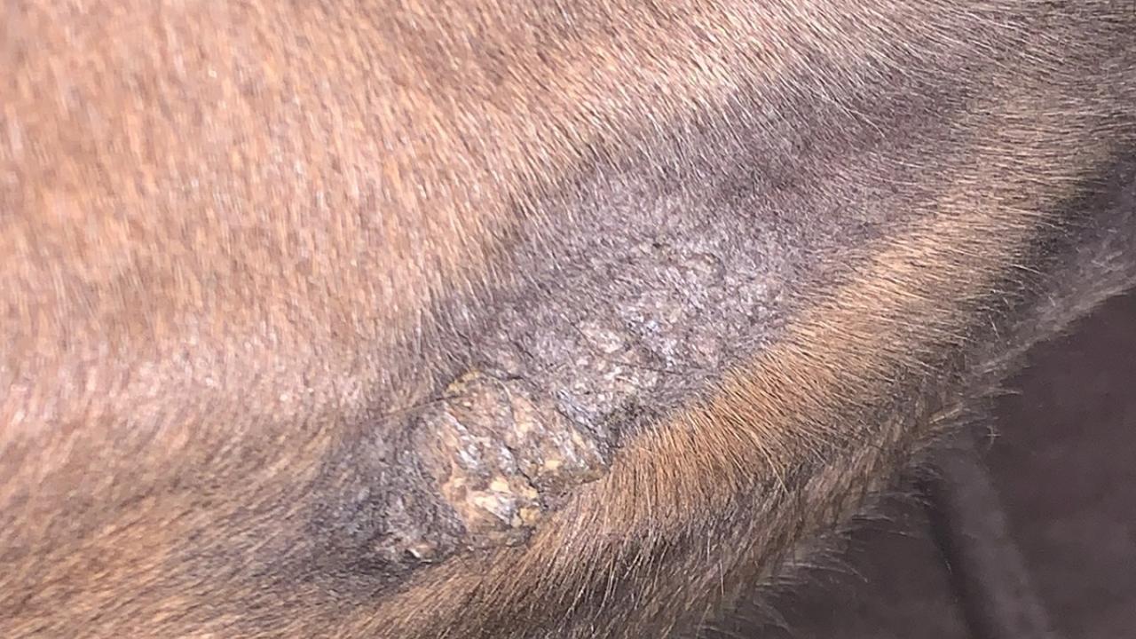
Fibroblastic Sarcoid — Pictures & Identification
Fibroblastic sarcoids are aggressive, looking like raw, fleshy masses. They ulcerate easily, oozing and bleeding with a wet, hemorrhagic surface. Roots extend deep, making them “proud flesh” mimics.

Sarcoids in horses: all you need to know | Horse & Hound’s vet library
Fibroblastic sarcoid on horse leg — equine sarcoid pictures identification.
Often on legs or wounds. Mistaken for granulation tissue or squamous cell carcinoma. However, carcinomas invade more destructively.
Sarcoids | School of Veterinary Medicine
Fibroblastic sarcoid on horse abdomen — equine sarcoid images.
Flies love them, so cover up.
Mixed Sarcoid — Pictures & Identification
Mixed sarcoids blend two or more types, like verrucous with nodular elements. They show warty patches alongside lumps or ulcers, often in clusters.
UK Vet Equine – Managing periocular sarcoids
Mixed sarcoid on horse eyelid — equine sarcoid pictures identification.
Anywhere, but common where types overlap. Confused with evolving single types. As a result, they indicate progression.
Sarcoids | School of Veterinary Medicine
Mixed sarcoid on horse neck — equine sarcoid images.
Monitor for changes.
Malevolent Sarcoid — Pictures & Identification
Malevolent sarcoids are rare but vicious, spreading like cords with nodules and ulcers. They infiltrate widely, resembling networks of aggressive growths.
Sarcoids | School of Veterinary Medicine
Malevolent sarcoid on horse groin — equine sarcoid pictures identification.
Often limbs or head. Mistaken for advanced fibroblastic, but more invasive. However, they don’t metastasize internally.
Example:
Sarcoids in horses: all you need to know | Horse & Hound’s vet library
Malevolent sarcoid on horse body — equine sarcoid images.
Tough to treat; act fast.
Sarcoid Pictures by Body Location
Location influences appearance and treatment. Here’s how sarcoids show up in common spots, with notes on care.
Sarcoid on horse leg: Most frequent, often fibroblastic or nodular. Legs endure trauma, so growths ulcerate easily. Treatment considers mobility — bandages help, but avoid constriction.
The Sinister Sarcoid – Springhill Equine Veterinary Clinic
Sarcoid on horse leg — horse sarcoid pictures.
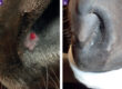
Sarcoid on horse ear: Verrucous or mixed, scaly and irritating. Ears twitch, worsening ulcers. Topical treatments work, but protect from flies.
Sarcoids | School of Veterinary Medicine
Sarcoid on horse ear — equine sarcoid images.
Sarcoid on horse eye/eyelid: Nodular or verrucous, risking vision. Swelling or discharge signals issues. Sensitive area demands gentle options.
UK Vet Equine – Managing periocular sarcoids
Sarcoid on horse eyelid — sarcoid identification pictures.
Sarcoid on horse belly/sheath: Occult or fibroblastic, hidden and moist. Sheath ones swell painfully. Cleanliness key for treatment.
Sarcoids in horses: all you need to know | Horse & Hound’s vet library
Sarcoid on horse sheath — equine sarcoid pictures.
What Else Could It Be?
Not every bump is a sarcoid. Here’s a comparison of common mimics:
- Melanoma: Gray horses prone; dark, nodular under tail or lips. Unlike sarcoids, they can metastasize. Biopsy differentiates.
- Proud flesh/granulation tissue: From wounds, exuberant pink growth. Heals with bandaging; sarcoids persist.
- Papillomas (warts): Viral in young horses, crusty on muzzle. Resolve naturally; sarcoids don’t.
- Fibroma: Benign, firm lumps. Less aggressive than nodular sarcoids.
- Squamous cell carcinoma: Ulcerated, invasive on light skin. Sun-related; biopsied for confirmation.
When to biopsy? If growth changes rapidly, ulcerates, or doesn’t respond to basic care. Vets recommend it for accurate diagnosis, especially before treatment. As per UC Davis guidelines, early biopsy prevents missteps.
Treatment Options
Sarcoids demand tailored approaches. Options include surgery, cryotherapy, laser removal, or topical creams. For a full rundown, check our guide on Sarcoid Growth on Horses: Understanding Treatment Options. Natural alternatives like bloodroot paste target roots without harsh chemicals. Our Veterinary Grade Bloodroot Paste (1oz) or Veterinary Grade Bloodroot Paste (2oz) applies easily for home use. See all options at All VetGrade Products. Success varies by type and location — occult respond well, malevolent less so. Consult your vet first.
Curious about timelines? Read How Long Does It Take for a Sarcoid to Fall Off?
For basics, start with What Is a Sarcoid on a Horse?
Ready to Tackle Sarcoids?
Armed with these equine sarcoid pictures, you’re set to monitor your horse effectively. Early action keeps minor issues from becoming major headaches. For more tips, subscribe for sarcoid treatment tips and exclusive savings. Stay proactive — your horse depends on it.
Frequently Asked Questions
Q: What does a sarcoid on a horse look like?
A: A sarcoid on a horse appears as a hairless patch, wart-like growth, nodule, or ulcerated mass depending on the type. Common features include gray, scaly skin or fleshy, bleeding surfaces. Always compare to equine sarcoid pictures and consult a vet for confirmation.
Q: How can I tell if my horse has a sarcoid?
A: Check for persistent bumps, hair loss, or warts that don’t heal, especially on legs, head, or groin. Use equine sarcoid images to match appearances, noting changes over time. A vet biopsy confirms it’s a sarcoid versus other conditions.
Q: What is the difference between a sarcoid and a wart on a horse?
A: Sarcoids are viral tumors that persist and can grow aggressively, while warts (papillomas) are benign and often resolve in young horses. Sarcoids lack the clustered, cauliflower look of warts. Biopsy distinguishes them accurately.
Q: Can a sarcoid be mistaken for melanoma?
A: Yes, sarcoids can mimic melanoma, especially nodular types on gray horses. Melanomas are darker and may metastasize, unlike sarcoids. Location and biopsy help differentiate — melanomas favor under the tail.
Q: Should I biopsy a suspected sarcoid?
A: Biopsy a suspected sarcoid if it grows, ulcerates, or doesn’t respond to monitoring. This confirms the diagnosis and rules out cancers like squamous cell carcinoma. Vets recommend it before treatment for best results.
Q: Do sarcoids change appearance over time?
A: Sarcoids often change from flat occult types to warty or ulcerated forms if traumatized. Monitoring with photos tracks evolution. Early intervention prevents aggressive shifts.